Medial Collateral Ligament (MCL) Injury

Typical Symptoms

Injuries to the medial collateral ligament (MCL) causes pain on the inner aspect of the knee together with swelling, limited movement and often difficulty weight bearing. In more serious cases, there can be instability of the knee that affects function.

What causes it?

The MCL sits on the inner aspect of the knee and is usually injured following a contact injury during sport where the lower leg is either pushed outwards or the knee is pushed inwards from a contact to the outer aspect.

Depending on the severity of the force, there can be injury to the deep or the superficial parts which can lead to stability issues with side-to-side movement or rotation.

How can I help myself?

It is useful to think back to the injury and how it happened; if it was from a contact and there is considerable swelling and limitation in movement, simple PRICE measures can help reduce this but care must be taken not to injure the skin.

If there is pain with walking or activity, offloading the knee with crutches can be helpful as can analgesia such as anti-inflammatories. A support or strapping can also help the knee feel more stable.

When to seek help?

If your symptoms are considerable, with pain, swelling and significantly impacted function, and there has been a considerable injury, you should be assessed as soon as possible.

What are the treatment options?

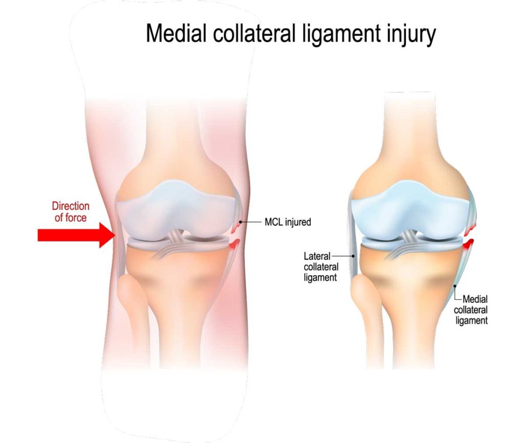
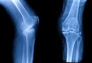
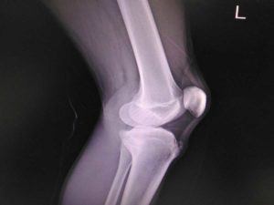
Your clinician will assess you with a thorough history and examination; once the location of pain is identified, they may obtain an initial X-ray of the knee to look for a bony injury such as an avulsion (where a section of bone is pulled off). In situations where an MCL injury is suspected, they may perform an ultrasound scan to look for swelling, tearing or inflammation.

If pain and swelling are significant, they may recommend bracing and crutches, but rehabilitation with a physiotherapist is key to recovery.

In situations where symptoms are ongoing, your clinician may discuss an ultrasound-guided injection of steroid or prolotherapy for inflammation or tearing. Usually this will be preceded by further imaging in the form of an MRI to gather more information. In some situations, surgical intervention may be required to repair the ligament.

Related treatments